Home
/ Chest Muscles Anatomy Labeled - Muscle Chest Anatomy Anatomy Drawing Diagram : It also protects several vital organs of the chest, such as the heart, aorta, vena cava, and.
Chest Muscles Anatomy Labeled - Muscle Chest Anatomy Anatomy Drawing Diagram : It also protects several vital organs of the chest, such as the heart, aorta, vena cava, and.
Chest Muscles Anatomy Labeled - Muscle Chest Anatomy Anatomy Drawing Diagram : It also protects several vital organs of the chest, such as the heart, aorta, vena cava, and.. Labeled muscles of lower leg. Dr calum worsley and assoc prof craig hacking et al. This all changes after hysterectomy. Several muscles that move the arms, head, and neck have their origins on the sternum. Except for the brain, the trunk houses all the vital organs of the human body.
This all changes after hysterectomy. Related to human arm muscles anatomy. Rib cage anatomy the rib cage, shaped in a mild cone shape and more flexible than most bone sets, is made up of varying elements such as the thoracic vertebra, 12 equally paired ribs, costal cartilage, and held together anteriorly by the sternum. The pectoralis major and the pectoralis minor, known collectively as your pecs. This might sound like a strange question, right?
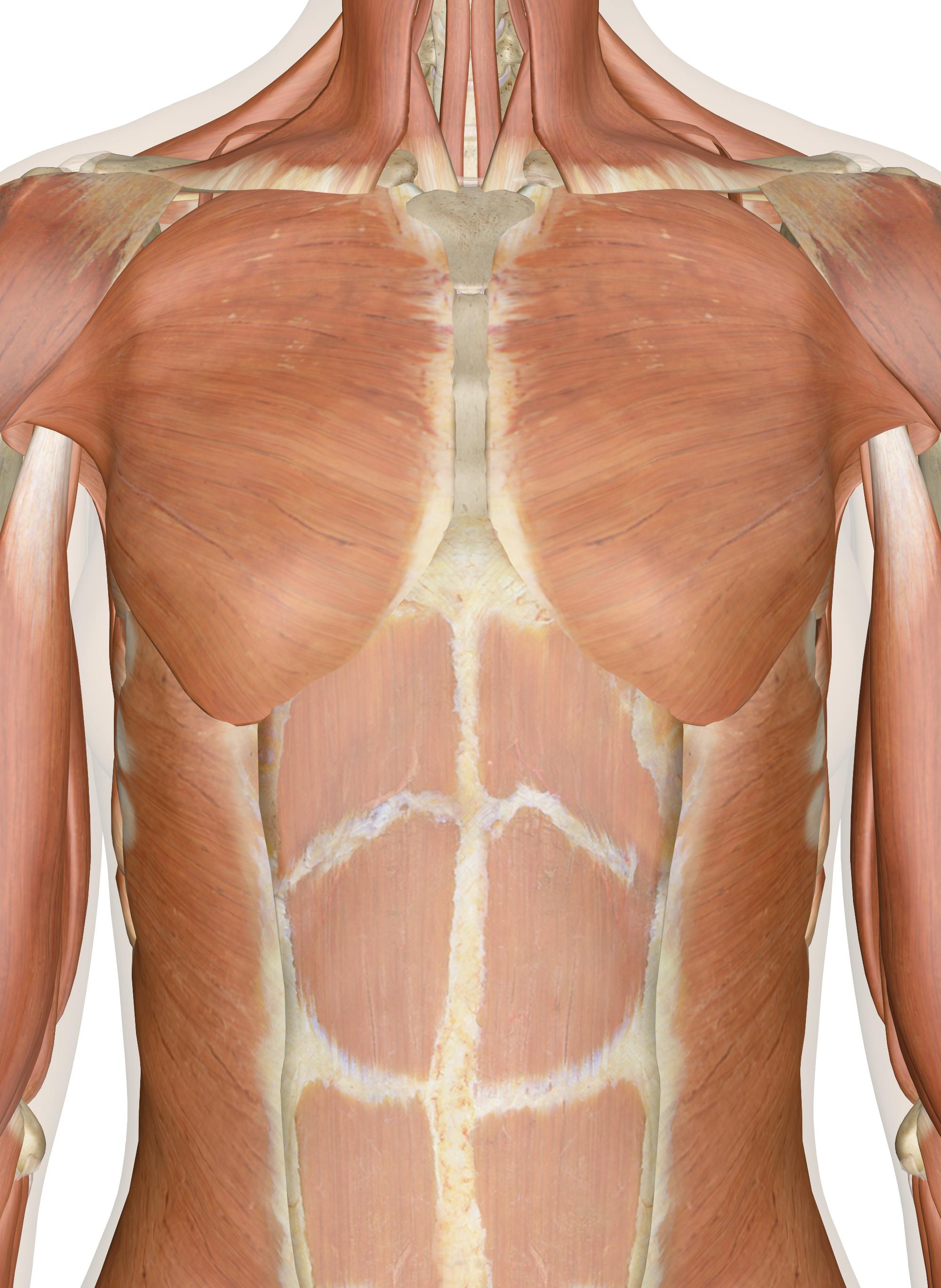
Muscles Of The Chest And Upper Back from innerbody.imgix.net Labeled muscles of lower leg. The circulatory system does most of its. Chest muscle anatomy the pectoralis major muscles also known as the pecs are located on the front of the rib cage and form the major muscles of the chest. Each of these muscles has its origin on the scapula and inserts around the head of the humerus. Except for the brain, the trunk houses all the vital organs of the human body. Several muscles that move the arms, head, and neck have their origins on the sternum. Arm muscle anatomy arm anatomy human body anatomy human anatomy and physiology anatomy study muscle diagram anatomy models muscular system bones and muscles. You go to the gym to train your abs.
Use the mouse scroll wheel to move the images up and down alternatively use the tiny arrows (>>) on both side of the image to move the images.>>) on both side of the image to move the images.
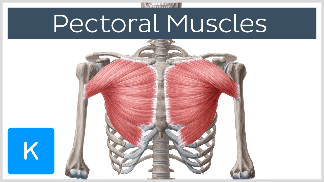
Here is the same image with the chest muscles labeled. Plus, how to target each to make them bigger and stronger. This muscle is divided into three named parts: Dr calum worsley and assoc prof craig hacking et al. The pectoralis major, pectoralis minor, serratus anterior and subclavius. Chest muscle anatomy the pectoralis major muscles also known as the pecs are located on the front of the rib cage and form the major muscles of the chest. When you think of abs, what muscle do you typically think of? Muscles and layers of the thoracic cavity. The pectoral region is located on the anterior chest wall. Short video of the chest muscles on the anterior torso identifies: Ct neck with annotated scrollable images. Rib cage anatomy the rib cage, shaped in a mild cone shape and more flexible than most bone sets, is made up of varying elements such as the thoracic vertebra, 12 equally paired ribs, costal cartilage, and held together anteriorly by the sternum. Each of these muscles has its origin on the scapula and inserts around the head of the humerus.
Rib cage anatomy the rib cage, shaped in a mild cone shape and more flexible than most bone sets, is made up of varying elements such as the thoracic vertebra, 12 equally paired ribs, costal cartilage, and held together anteriorly by the sternum. Related to human arm muscles anatomy. Use the mouse scroll wheel to move the images up and down alternatively use the tiny arrows (>>) on both side of the image to move the images.>>) on both side of the image to move the images. The anterior serratus pulls the scapula outward which lifts the shoulder. Short video of the chest muscles on the anterior torso identifies:
Pectoral Muscles Area Innervation Function Human Anatomy Kenhub Youtube from i.ytimg.com This article lists a series of labeled imaging anatomy cases by system and modality. Chest muscle anatomy the pectoralis major muscles also known as the pecs are located on the front of the rib cage and form the major muscles of the chest. Male shoulder and chest muscles labeled chart on white labeled human anatomy diagram of male shoulder, biceps, arm, and chest muscles frontal anterior view on a white background. When you think of abs, what muscle do you typically think of? Dr calum worsley and assoc prof craig hacking et al. Human anatomy for muscle, reproductive, and skeleton. The sternum, commonly known as the breastbone, is a long, narrow flat bone that serves as the keystone of the rib cage and stabilizes the thoracic skeleton. Muscle anatomy body anatomy anatomy study muscular system human anatomy and physiology back muscles shoulder muscles massage therapy physical therapy.
This article lists a series of labeled imaging anatomy cases by system and modality.
(1) the pectoralis major, and (2) the pectoralis minor. The pectoralis major, pectoralis minor, serratus anterior and subclavius. Muscle anatomy labeled 12 photos of the muscle anatomy labeled muscle anatomy diagrams, muscle anatomy labeling exercises, muscle anatomy labeling worksheet, muscle anatomy labelling quiz, muscle models anatomy. It also protects several vital organs of the chest, such as the heart, aorta, vena cava, and. The torso muscles attach to the skeletal core of the trunk, and depending on their location are divided into two large groups: Serratus anterior superior, serratus anterior intermediate, serratus anterior inferior and runs from the front of the chest around the side to the scapula. This muscle is divided into three named parts: When you think of abs, what muscle do you typically think of? This tool was chosen to show segmental lung anatomy (international and ikeda). Short video of the chest muscles on the anterior torso identifies: Muscle anatomy exercise chart 12 photos of the muscle anatomy exercise chart muscle anatomy exercise chart, human muscles, muscle anatomy exercise chart. Use the mouse scroll wheel to move the images up and down alternatively use the tiny arrows (>>) on both side of the image to move the images.>>) on both side of the image to move the images. The anatomy of your abdominal muscles.
This article lists a series of labeled imaging anatomy cases by system and modality. Each of these muscles has its origin on the scapula and inserts around the head of the humerus. Human anatomy for muscle, reproductive, and skeleton. This mri chest (thorax) axial cross sectional anatomy tool is absolutely free to use. As the cursor is moved over an anatomical area of the lung parenchyma, the segment is highlighted and labeled:
Thorax Wikipedia from upload.wikimedia.org The sternum, commonly known as the breastbone, is a long, narrow flat bone that serves as the keystone of the rib cage and stabilizes the thoracic skeleton. It contains four muscles that exert a force on the upper limb: Ventral trunk muscles (overview) the trunk (torso) is the central part of the body to which the head and the limbs are attached. Browse 20 labeled arm muscles stock photos and images available, or start a new search to explore more stock photos and images. Human anatomy for muscle, reproductive, and skeleton. Über 7 millionen englischsprachige bücher. The tendons of these muscles surround and support the humerus while the contraction of the muscles rotates, adducts, or abducts the humerus. This tool was chosen to show segmental lung anatomy (international and ikeda).
Each of these muscles has its origin on the scapula and inserts around the head of the humerus.
Ventral trunk muscles (overview) the trunk (torso) is the central part of the body to which the head and the limbs are attached. Rib cage anatomy, labeled vector illustration diagram. This tool was chosen to show segmental lung anatomy (international and ikeda). Here is the same image with the chest muscles labeled. The rotator cuff consists of four muscles: As the cursor is moved over an anatomical area of the lung parenchyma, the segment is highlighted and labeled: Supraspinatus, infraspinatus, subscapularis, and teres minor. The circulatory system does most of its. (1) the pectoralis major, and (2) the pectoralis minor. Here, we break down the anatomy of your chest muscles. It contains four muscles that exert a force on the upper limb: Folge deiner leidenschaft bei ebay! Arm muscle anatomy arm anatomy human body anatomy human anatomy and physiology anatomy study muscle diagram anatomy models muscular system bones and muscles.
Coronal c+ portal venous phase chest muscles anatomy. Rib cage anatomy the rib cage, shaped in a mild cone shape and more flexible than most bone sets, is made up of varying elements such as the thoracic vertebra, 12 equally paired ribs, costal cartilage, and held together anteriorly by the sternum.